Structure and Function of Heart
The heart is a triangular-shaped, hollow, muscular organ. It is situated on the left side between the two lungs. It consists of special involuntary muscles.
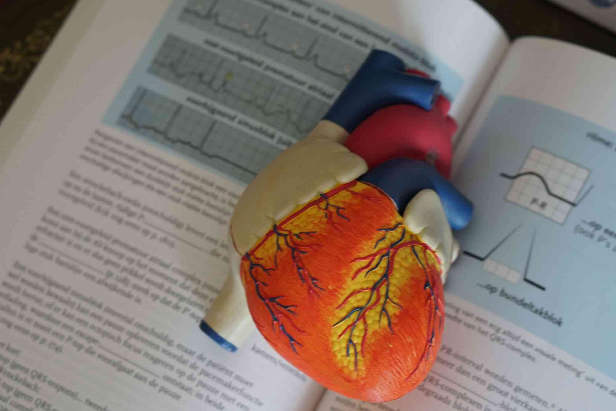
The heart is a triangular-shaped, hollow, muscular organ. It is situated on the left side between the two lungs. It consists of special involuntary muscles. It is surrounded by a thin membrane named the pericardium. The heart of the wall consists of three layers:
- Epicardium: This consists of connective tissue. This layer is covered with epithelial tissue. Fat bodies are scattered on it.
- Myocardium: This layer is in between the epicardium and endocardium. It consists of strong involuntary muscles.
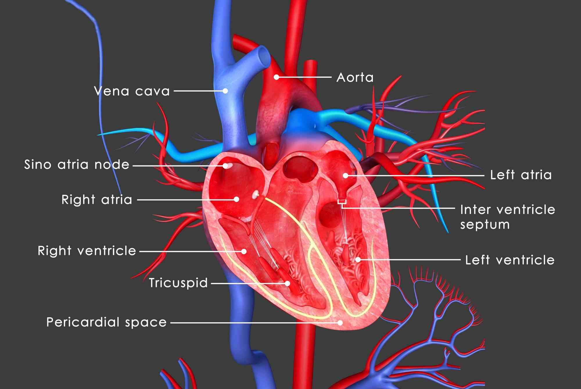
- Endocardium: This is the innermost layer. The chambers of the heart are surrounded by the endocardium. This layer also covers the valves. The inner part of the heart is hollow and four-chambered. The upper chambers are known as the right and the left auricle. The lower chambers are the right ventricle and the left ventricle. The two atria and ventricles are separated by inter auricular and the inter-ventricular septum, respectively.
(Article continues below)
Valves guard the aperture between the two atria (singular atrium) and ventricles. The right auriculo-ventricular aperture guarded by a tricuspid by a bicuspid valve made up of two flaps. The opening of the aorta and the pulmonary artery is guarded by valves called semilunar valves, which allow the transport of blood in the direction and prevents the backflow of blood.
Circulation of blood through the heart
The heart acts as a pump. The heart works by contraction and relaxation. The continuous contraction and relaxation transport blood throughout the whole body.
The contraction of the heart is called systole, and the relaxation of the heart is called the diastole. A complete contraction(systole) and relaxation(diastole) of the heart constitutes a heartbeat. Due to the relaxation of atria(auricles) the blood enters the heart from different parts of the body. Such as -deoxygenated blood from the superior vena cava enters the right atrium. At the same time, the oxygenated blood enters the left atrium through the pulmonary veins from the lungs.
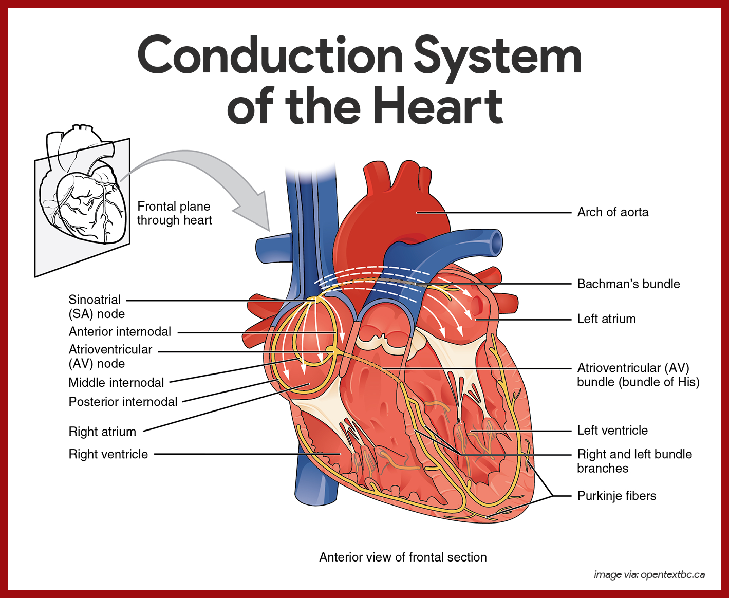
The walls of the two atriums contact and then the muscles of the ventricle relax. As a result, the tricuspid valve is situated between the right Sino auricular ventricular apertures open. So, the deoxygenated blood from the right auricle enters the right ventricle. At the same time, the left Sino auricular ventricular aperture, guarded by the bicuspid valve, opens. The oxygenated blood enters the left ventricle from the left auricle. During this period, the left and the right auriculoventricular apertures are closed by their tricuspid and bicuspid valves. So, the blood of the ventricle cannot return to the atrium.
When the two ventricles relax, deoxygenated blood from the right ventricle passes through the aorta towards the body, and the opening of both the arteries (aorta and pulmonary artery) are closed by the semilunar valves which prevent blood from returning into the ventricle. Thus, successive contraction and relaxation of the atrium and ventricle help in the continuous transportation of blood.
Function of Heart
The heart is the principal organ of the circulatory system. It helps blood to keep moving. The human heart is divided into four chambers. In higher animals, the chambers are completely separated. So, oxygenated and deoxygenated blood do not mix.
Recommended reading:
Source:
Anatomy Physiology The Unity of Form and Function, Eighth Edition (KENNETH S. SALADIN)


